
East Valley Ophthalmology provides this retina glossary to enhance understanding of eye care and surgery terms. This resource defines common medical terminology,
aiding comprehension of diagnoses and treatments related to the retina.
Purpose of the Glossary
East Valley Ophthalmology’s retina glossary serves a crucial role in patient education and empowerment. The primary purpose is to demystify complex medical terminology associated with the retina, its functions, and potential conditions. This glossary aims to bridge the communication gap between eye care professionals and individuals seeking to understand their eye health.
By providing clear and concise definitions for terms like aberration, astigmatism, and conditions such as retinal detachment and macular degeneration, the glossary enables patients to actively participate in their care. Understanding these terms fosters informed decision-making regarding treatment options and promotes a collaborative relationship with their ophthalmologist. It’s designed to be a readily accessible resource, supporting a deeper comprehension of diagnoses and the rationale behind recommended interventions, ultimately improving the overall patient experience.
Target Audience
This retina glossary from East Valley Ophthalmology is designed for a broad audience, encompassing anyone interested in learning more about the retina and related eye conditions. Primarily, it’s intended for patients who have been diagnosed with a retinal issue, such as diabetic retinopathy or require understanding of procedures like a fundus examination.
The glossary also benefits individuals proactively seeking knowledge about eye health and preventative care. Family members and caregivers supporting loved ones with retinal conditions will find it a valuable resource. Furthermore, students and those with a general interest in anatomy and physiology – specifically the layers of the retina and cell bodies and synapses – can utilize this glossary to expand their understanding. No prior medical knowledge is assumed, ensuring accessibility for all seeking clear definitions of essential eye care terminology.

Anatomy of the Retina
Associate Professor Trevor Sherwin’s research details the retina’s structure, including a sagittal section, cellular detail, and its three layers of cell bodies and two synaptic layers.
Layers of the Retina
The retina, a complex neural tissue, is meticulously organized into distinct layers, each playing a crucial role in vision. These layers, as detailed in research by Associate Professor Trevor Sherwin, facilitate the conversion of light into neural signals.
Fundamentally, all vertebrate retinas share a common architecture: three layers of cell bodies and two layers of synapses. The outermost layer contains photoreceptors – rods and cones – responsible for detecting light. Beneath this lies the outer plexiform layer, where photoreceptors synapse with bipolar cells.
The inner nuclear layer houses the cell bodies of bipolar, amacrine, and horizontal cells, contributing to signal processing. Following this is the inner plexiform layer, where bipolar cells synapse with ganglion cells. Finally, the ganglion cell layer contains the output neurons of the retina, whose axons form the optic nerve, transmitting visual information to the brain. Understanding these layers is fundamental to comprehending retinal function and pathology.
Cell Bodies and Synapses
Within the retina’s layered structure, cell bodies and synapses orchestrate the intricate process of visual signal transduction. As highlighted by Associate Professor Trevor Sherwin’s research, these components are not randomly distributed but are precisely arranged to optimize visual processing.
Photoreceptors, bipolar cells, amacrine cells, horizontal cells, and ganglion cells each contribute unique cell bodies to specific retinal layers. These cells communicate via synapses – specialized junctions where signals are transmitted. The outer plexiform layer is characterized by synapses between photoreceptors and bipolar cells, initiating signal processing.
The inner plexiform layer features synapses between bipolar and ganglion cells, refining the signal before transmission to the brain. Amacrine cells modulate these signals laterally. This complex interplay of cell bodies and synapses ensures efficient and accurate conversion of light into neural impulses, ultimately enabling vision.
Sagittal Section of the Retina
A sagittal section of the retina, as studied by Associate Professor Trevor Sherwin, provides a crucial cross-sectional view of its layered architecture. This perspective reveals the distinct arrangement of photoreceptors, cell bodies, and synaptic layers, essential for understanding retinal function.
Examining this section demonstrates how light traverses the different layers – from the outer retina containing photoreceptors, through the outer plexiform layer where initial signal processing occurs, to the inner retina with bipolar and ganglion cells.
The sagittal view clarifies the spatial relationships between these components, highlighting how signals are relayed and refined. It visually demonstrates the organization that allows for efficient conversion of light into neural signals. Understanding this anatomy is fundamental to diagnosing and treating various retinal conditions, as detailed in the East Valley Ophthalmology glossary.
Cellular Detail of the Retina
Detailed examination, as highlighted by Associate Professor Trevor Sherwin’s research, reveals the intricate cellular composition of the retina. All vertebrate retinas share a common structure: three layers of cell bodies – photoreceptors, bipolar cells, and ganglion cells – interspersed with two layers of synapses.
Photoreceptors, including rods and cones, initiate the visual process by converting light into electrical signals. These signals are then transmitted to bipolar cells, which relay information to ganglion cells. The axons of ganglion cells form the optic nerve, carrying visual information to the brain.
The East Valley Ophthalmology glossary emphasizes understanding these cellular interactions. The precise arrangement and function of these cells are critical for visual acuity and perception. Studying this cellular detail is vital for comprehending retinal diseases and developing effective treatments.
Retinal Terminology ⎼ General
East Valley Ophthalmology’s glossary defines key terms like aberration, astigmatism, convergence, and focus point, crucial for understanding how light interacts with the retina.
Aberration
Within the realm of optics and ophthalmology, an aberration refers to a distortion or imperfection in the image formed by a lens or mirror – in this case, the eye’s optical system. These distortions prevent light rays entering the eye from converging to a single, clear focus point on the retina.
Specifically, aberrations are often related to astigmatism, a common refractive error where the cornea (the clear front surface of the eye) has an irregular shape. This irregular shape causes light to focus at multiple points instead of a single point, resulting in blurred or distorted vision. Different types of aberrations exist, impacting visual clarity in various ways.
Understanding aberrations is vital for diagnosing and correcting vision problems. Corrective lenses, such as glasses or contact lenses, are designed to counteract these distortions and restore clear vision by properly refracting light onto the retina. Advanced diagnostic tools help ophthalmologists identify and measure aberrations to personalize treatment plans.
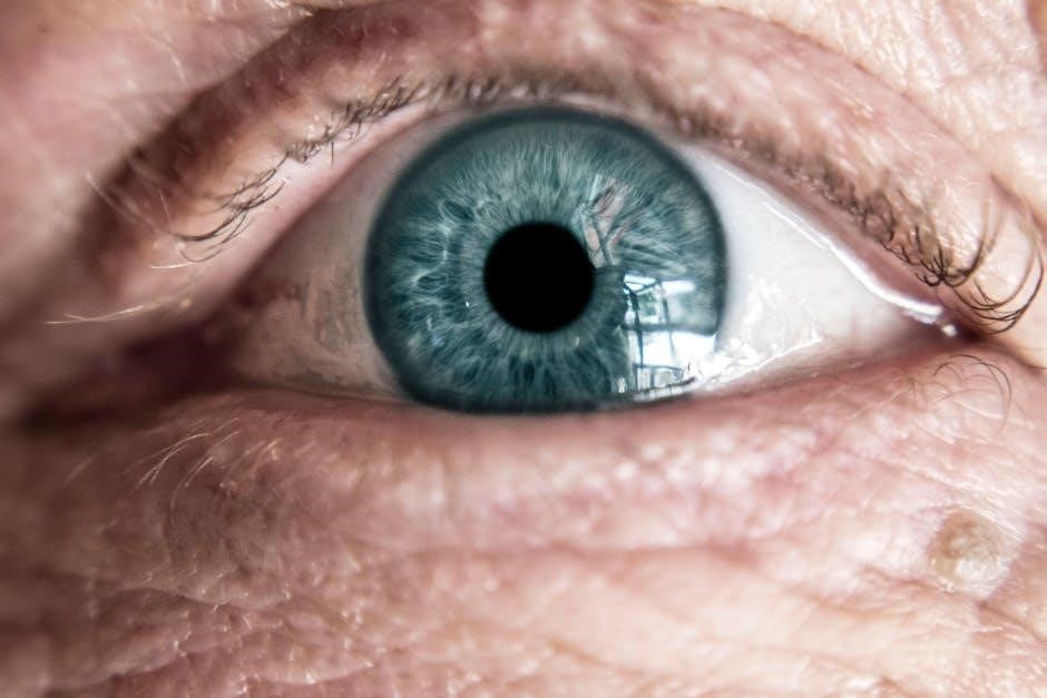
Astigmatism
Astigmatism is a common vision condition characterized by an irregular curvature of the cornea – the clear front surface of the eye – or, less frequently, the lens. This irregular shape prevents light from focusing precisely on a single point on the retina, leading to blurred or distorted vision at all distances.
Unlike nearsightedness or farsightedness, astigmatism doesn’t necessarily mean you see blurry at a specific distance; it causes distortion regardless. It often occurs alongside other refractive errors. Aberrations, distortions in how light enters the eye, are closely linked to astigmatism, as the irregular shape causes the inability of light rays to converge properly.
Astigmatism is easily corrected with corrective lenses, including glasses and contact lenses, which compensate for the irregular curvature. Surgical options, like LASIK, can also permanently reshape the cornea to correct astigmatism. Regular eye exams are crucial for detecting and managing this condition.
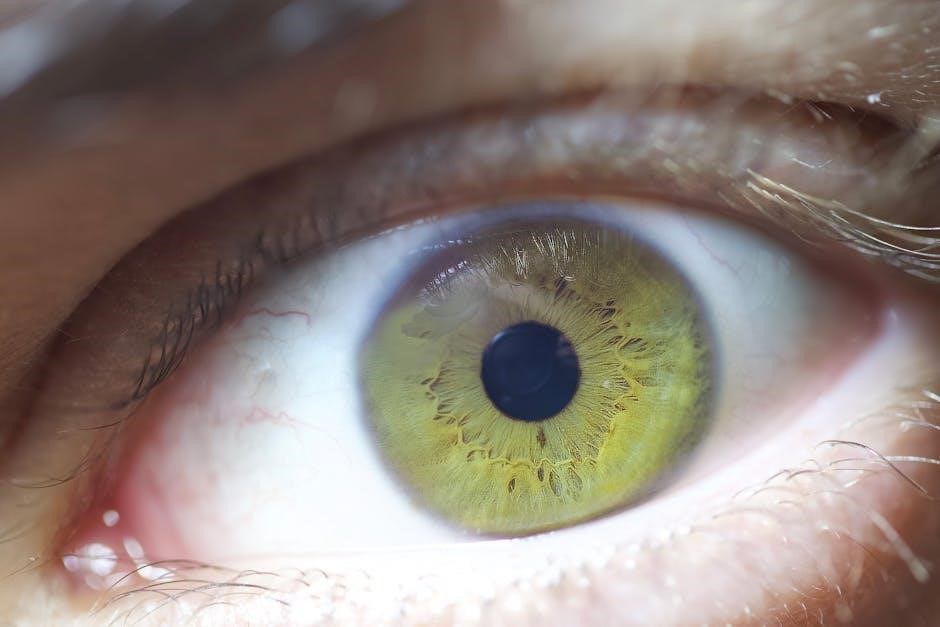
Convergence (of light rays)
Convergence, in the context of vision, refers to the process by which the eye’s lens bends incoming light rays so they precisely meet at a single focus point on the retina. This precise focusing is essential for clear, sharp vision. The cornea and lens work together to refract, or bend, light;
When light rays don’t converge correctly – often due to refractive errors like nearsightedness, farsightedness, or astigmatism – the image projected onto the retina becomes blurred. Aberrations can also disrupt this process, causing distortions that prevent proper convergence.
The ability of the eye to converge light effectively depends on the shape of the cornea and lens, as well as the eye’s focusing muscles. Corrective lenses, such as glasses or contacts, are designed to help redirect light rays for proper convergence. Maintaining healthy eye function is vital for optimal visual acuity and clear sight.
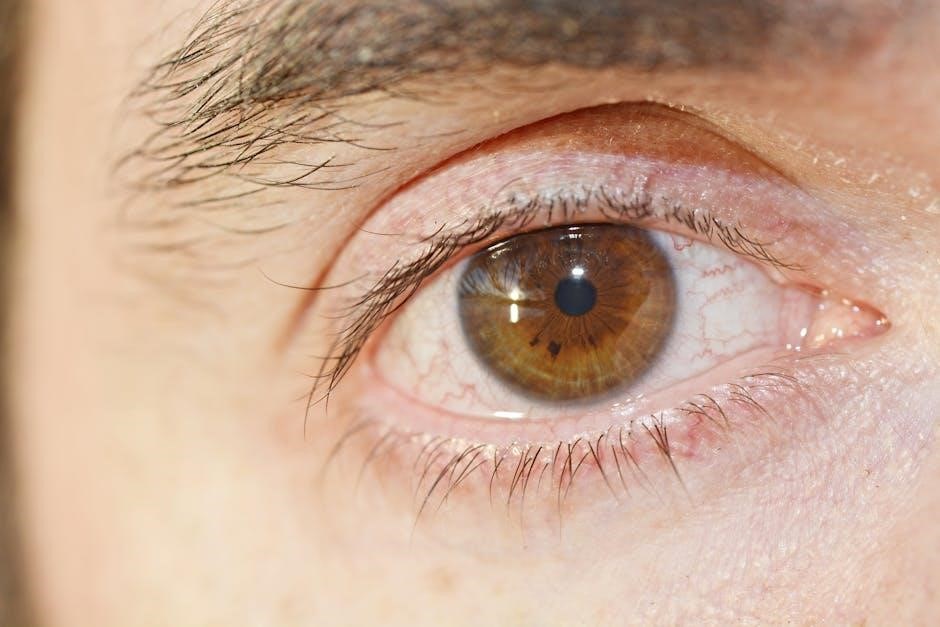
Focus Point
The focus point, crucial for clear vision, is the precise location on the retina where light rays converge after passing through the cornea and lens of the eye; Ideally, this point creates a sharp, detailed image. Achieving accurate focus relies on the eye’s ability to refract, or bend, light effectively.
Distortions, known as aberrations, or refractive errors like astigmatism, can prevent light from converging at a single, defined focus point, resulting in blurred vision. Proper convergence of light rays is therefore paramount.
The retina’s photoreceptor cells, responsible for detecting light, are concentrated around the fovea, the central focus point. This area provides the highest visual acuity. Any deviation from precise focusing compromises image clarity. Corrective lenses aim to shift the focus point directly onto the retina, restoring sharp vision and optimal visual performance.

Specific Retinal Conditions & Terms
This section details common retinal issues like retinal detachment, macular degeneration, diabetic retinopathy, and retinal tears, offering clarity on these conditions.
Retinal Detachment
Retinal detachment occurs when the retina separates from the underlying tissue in the back of the eye. This separation can lead to vision loss if not promptly treated. It often begins with a tear or hole in the retina, allowing fluid to accumulate beneath it, pushing it away from its supporting layers.
Symptoms can include a sudden increase in floaters – small specks or clouds moving in your field of vision – flashes of light, or a shadow appearing in your peripheral (side) vision. The experience is often described as a curtain coming down over one’s vision. Prompt diagnosis and treatment, often involving laser surgery or a vitrectomy, are crucial to reattach the retina and preserve sight.
Understanding the urgency of seeking medical attention is paramount when experiencing these symptoms, as delays can significantly impact the potential for successful intervention and long-term visual outcomes. Early detection dramatically improves the chances of restoring retinal function.
Macular Degeneration
Macular degeneration is a common age-related condition affecting the central part of the retina, known as the macula. This area is responsible for sharp, central vision needed for tasks like reading and driving. There are two main types: dry and wet.
Dry macular degeneration is more prevalent and progresses slowly, causing gradual vision loss. Wet macular degeneration, while less common, is more aggressive, involving abnormal blood vessel growth under the retina which can lead to rapid central vision loss. Symptoms include blurred or distorted central vision, difficulty recognizing faces, and needing brighter light for reading.
While there’s no cure, treatments like anti-VEGF injections for wet AMD can slow progression and, in some cases, improve vision. Lifestyle modifications and nutritional supplements may help manage dry AMD. Regular eye exams are vital for early detection and intervention.
Diabetic Retinopathy
Diabetic retinopathy is a diabetes-related complication impacting the blood vessels of the retina. Prolonged high blood sugar levels can damage these vessels, leading to leakage, swelling, and ultimately, vision loss. It’s a leading cause of blindness in adults.
The condition progresses through stages, starting with non-proliferative diabetic retinopathy (NPDR), characterized by minor blood vessel changes. As it advances to proliferative diabetic retinopathy (PDR), abnormal new blood vessels grow on the retina, increasing the risk of bleeding and retinal detachment.
Early detection through regular dilated eye exams is crucial. Treatment options include laser therapy to seal leaking vessels, injections of anti-VEGF medications to reduce blood vessel growth, and, in severe cases, vitrectomy surgery. Strict blood sugar control, blood pressure management, and cholesterol monitoring are vital for prevention and management.
Retinal Tears
Retinal tears occur when the vitreous, the gel-like substance filling the eye, pulls on the retina, creating a tear. This often happens as people age and the vitreous naturally shrinks and pulls away from the retina – a common occurrence, but not always problematic.
However, if a tear develops, fluid can seep underneath the retina, potentially leading to a retinal detachment, a serious condition requiring immediate medical attention. Symptoms of a retinal tear can include sudden flashes of light (photopsia), a sudden increase in floaters, or a shadow in your peripheral vision.
Treatment typically involves laser photocoagulation or cryopexy (freezing) to seal the tear and prevent fluid from accumulating. Prompt diagnosis and treatment are essential to minimize the risk of retinal detachment and preserve vision. Regular eye exams are crucial for early detection.

Diagnostic Procedures
Diagnostic procedures, like OCT, retinal photography, and a thorough fundus examination, are vital for assessing retinal health and identifying potential issues effectively.
Optical Coherence Tomography (OCT)
Optical Coherence Tomography (OCT) is a non-invasive imaging technique providing high-resolution, cross-sectional views of the retina. This advanced diagnostic tool utilizes light waves to capture detailed images of the retinal layers, allowing ophthalmologists to visualize structures not easily seen during a standard fundus examination.
OCT is crucial for diagnosing and monitoring various retinal conditions, including macular degeneration, diabetic retinopathy, and retinal detachment. It precisely measures the thickness of retinal layers, detects fluid accumulation, and identifies subtle structural changes indicative of disease progression. The technology enables early detection and timely intervention, potentially preserving vision.
Furthermore, OCT assists in evaluating the effectiveness of treatments and guiding clinical decision-making. Its ability to provide quantitative data enhances diagnostic accuracy and facilitates personalized patient care. The detailed images generated by OCT are invaluable for understanding the complex anatomy and pathology of the retina.
Retinal Photography
Retinal Photography, also known as fundus photography, is a vital diagnostic technique capturing detailed images of the retina, optic disc, and blood vessels. This non-invasive procedure utilizes a specialized camera with a bright light source to visualize the back of the eye, providing a comprehensive record of its structural features.
Retinal photographs serve as a baseline for monitoring changes over time, particularly in conditions like diabetic retinopathy, glaucoma, and macular degeneration. They allow ophthalmologists to detect early signs of disease, track progression, and assess the effectiveness of treatment interventions. The images document abnormalities such as hemorrhages, exudates, and optic nerve damage.
Furthermore, retinal photography facilitates communication and collaboration among eye care professionals. High-resolution images can be easily shared for second opinions or remote consultations. This valuable diagnostic tool plays a crucial role in preserving vision and ensuring optimal patient outcomes.
Fundus Examination
A Fundus Examination is a crucial part of a comprehensive eye exam, allowing the doctor to directly view the internal structures of the eye, including the retina, optic disc, macula, and blood vessels. This examination is performed using an ophthalmoscope, an instrument that shines a bright light into the eye;
During a fundus exam, the ophthalmologist assesses the health of these structures, looking for any signs of disease or abnormalities. This includes detecting conditions like retinal detachments, macular degeneration, diabetic retinopathy, and glaucoma. The examination reveals details about blood vessel health, optic nerve appearance, and the presence of any lesions or hemorrhages.
The fundus examination is a relatively quick and painless procedure, providing invaluable information for diagnosing and managing a wide range of eye conditions. It’s a cornerstone of preventative eye care and early disease detection, contributing significantly to preserving vision.

Video Editing Tools for Visual Aids (Related to Retina Education)
Microsoft Clipchamp empowers educators to create professional videos for retina education, utilizing easy recording, templates, and innovative AI editing features for clarity.
Microsoft Clipchamp Overview
Microsoft Clipchamp stands as a remarkably beginner-friendly and accessible video editor, designed to empower individuals with varying levels of experience to craft compelling videos. It’s a versatile tool perfect for educators aiming to visually explain complex retinal concepts, or for creating supplementary materials to accompany a retina glossary.
Unlike traditional, complex video editing software, Clipchamp operates directly within a web browser, eliminating the need for downloads or installations. This accessibility is a significant advantage, allowing users to create and edit videos from virtually any device with an internet connection. The platform boasts a robust suite of powerful editing tools alongside a diverse library of pre-designed video templates, streamlining the video creation process.
Whether you’re a creator, gamer, educator, or a professional, Clipchamp offers the features needed to produce high-quality, professional-looking videos quickly and efficiently. It’s a fantastic resource for bringing visual clarity to intricate medical topics like those found within a detailed retina glossary.
AI Editing Features in Clipchamp
Clipchamp distinguishes itself through its integration of cutting-edge Artificial Intelligence (AI) editing features, significantly streamlining the video creation workflow – particularly useful when illustrating concepts from a retina glossary. One standout feature is the AI-powered Silence Removal tool. This intelligently identifies and highlights silences exceeding three seconds, allowing for quick previewing and removal, resulting in a more concise and engaging final product.
Beyond silence detection, Clipchamp offers AI transcript-based editing. This innovative functionality enables users to edit videos directly from the automatically generated transcript, accelerating the editing process and ensuring accuracy. These AI tools are invaluable for educators or medical professionals creating visual aids to explain complex retinal anatomy or conditions detailed in a comprehensive glossary.
By automating tedious tasks, Clipchamp’s AI features empower users to focus on the creative aspects of video production, ultimately enhancing the clarity and impact of their educational content.
Clipchamp’s Silence Removal Tool
Clipchamp’s AI Silence Removal tool is a game-changer for creating polished educational videos, especially when explaining intricate details from a retina glossary. This feature automatically scans video timelines, pinpointing silences lasting longer than approximately three seconds. It doesn’t simply delete them; it highlights each potential gap, allowing for careful previewing before removal.
Users have the flexibility to remove silences individually or utilize batch removal for a faster workflow. This is incredibly beneficial when producing instructional content on retinal anatomy, conditions like macular degeneration, or diagnostic procedures. Eliminating unnecessary pauses improves viewer engagement and comprehension.
The tool then exports a tightened video timeline, resulting in a more dynamic and professional presentation. This ensures that crucial information from the retina glossary is delivered concisely and effectively, maximizing the impact of the visual aid.
Easy Editing Features in Clipchamp
Clipchamp’s online video editor offers a suite of essential, user-friendly tools perfect for crafting clear and concise visual aids based on a retina glossary. Creators can precisely cut, trim, crop, and rotate footage, ensuring a professional presentation of complex anatomical diagrams or diagnostic imagery like OCT scans.
Splitting clips allows for focused explanations of specific terms – such as retinal detachment or aberrations – while zooming features highlight crucial details within fundus photographs. Adjusting video speed can slow down fast-paced explanations of cellular detail, aiding comprehension.
Furthermore, adding or removing audio, filters, and transitions enhances visual appeal and clarity. These features empower educators and medical professionals to transform a simple glossary into an engaging and informative video resource, improving understanding of retinal health.

Resources & Further Learning
East Valley Ophthalmology and Associate Professor Trevor Sherwin’s research offer valuable insights into retinal anatomy and conditions, complementing this retina glossary.
East Valley Ophthalmology
East Valley Ophthalmology, a leading eye care provider in Mesa, Arizona, is dedicated to delivering exceptional vision care services. They offer a comprehensive retina glossary as a valuable resource for patients seeking to understand complex eye-related medical terminology. This glossary aims to demystify the language surrounding eye care and eye surgery, empowering individuals to actively participate in their treatment journey.

Understanding terms like “aberration” and “astigmatism,” which impact how light converges on the retina, is crucial for grasping potential vision issues. East Valley Ophthalmology’s commitment extends beyond treatment; they prioritize patient education, ensuring individuals feel informed and confident about their ocular health. Their team of skilled eye doctors provides personalized care, utilizing advanced diagnostic tools and techniques to address a wide range of retinal conditions.
By offering this accessible retina glossary, East Valley Ophthalmology reinforces its dedication to patient-centered care and promotes a deeper understanding of eye health within the community.
Associate Professor Trevor Sherwin’s Research
Associate Professor Trevor Sherwin’s research delves into the intricate anatomy and physiology of the retina, providing foundational knowledge for understanding retinal function and disease. His work meticulously examines the layers of the retina, detailing the arrangement of cell bodies and synapses – essential components for visual processing.
Sherwin’s investigations include detailed sagittal sections of the retina and high-resolution analysis of cellular detail, offering crucial insights into the complex neural circuitry. This research extends to comparative studies, notably examining the octopus retina, to broaden our understanding of visual systems across species. His findings contribute significantly to the development of effective treatments for retinal disorders.
Understanding these anatomical structures is vital when interpreting a retina glossary, as many terms relate directly to these layers and cellular components. Sherwin’s research provides a scientific basis for comprehending the definitions and implications of various retinal conditions.